
The Bone Bank: Strong Bones, Strong Future
When we talk about perimenopause we first think hot flashes, disrupted sleep, weight changes, and brain fog. These are the changes we notice because they affect how we feel every day. But something else is happening beneath the surface, often without any symptoms at all. Our bones may be getting weaker.
Most of us don't think much about our bones until we break one, develop back pain, or receive an unexpected diagnosis of osteopenia or osteoporosis. Yet bone health is fundamental to maintaining our independence and physical capabilities as we age. Strong bones allow us to hike, dance, travel, lift our grandchildren, enjoy sex, and continue doing the things we love.
Osteoporosis is far more serious than simply having thin bones. Fragility fractures can lead to chronic pain, loss of mobility, disability, and loss of independence. Hip fractures are particularly devastating. Approximately one in five older adults dies within a year of a hip fracture, although estimates vary by age, sex, and overall health. This does not mean the fracture directly causes every death, but the injury, surgery, immobility, and complications can accelerate a decline in someone who was previously functioning independently. Women experience the majority of osteoporotic fractures because they live longer and experience accelerated bone loss with menopause.
What concerns me is that we often wait until women are 65 to routinely screen for osteoporosis, even though the processes that weaken our bones begin decades earlier. By the time we discover significant bone loss, we may have missed years of opportunities to prevent it.
Bone Health Begins Long Before Menopause
Our bones are living tissue, constantly being broken down and rebuilt through a process called remodeling. Osteoclasts remove older bone, while osteoblasts build new bone. During childhood and adolescence, bone formation exceeds breakdown, and we accumulate most of our peak bone mass by our late twenties or early thirties.
Think of peak bone mass as the savings account you build early in life. Genetics, nutrition, physical activity, hormones, and overall health determine how much you accumulate. Once you reach peak bone mass, maintaining that reserve becomes increasingly important.
Gradual age-related bone loss can begin in our thirties and forties, although the timing and rate vary. During the late reproductive years, some women begin experiencing subtle changes in bone metabolism even before their final menstrual period. Bone loss accelerates most dramatically during the late perimenopausal years and early postmenopause, when estrogen levels decline substantially.
Estrogen helps regulate the balance between bone breakdown and formation by suppressing excessive osteoclast activity. When estrogen declines, bone resorption increases, and bone formation cannot always keep up. Women may lose approximately 10% of bone mineral density around the menopause transition, with some losing considerably more. Estimates approaching 20% over five to seven years represent the higher end rather than what every woman should expect.
Progesterone also has receptors and biological activity in bone tissue, and laboratory research suggests it may influence osteoblast function. However, the clinical evidence that progesterone alone prevents fractures or meaningfully preserves bone density is much weaker than the evidence for estrogen. I don't prescribe progesterone by itself as an osteoporosis-prevention treatment.
This is why I want women thinking about bone health well before their sixties. Menopause is a period of accelerated change, but the foundation for skeletal health is established much earlier.
Birth Control, Fertility Treatments, and Pregnancy
Our reproductive history can provide important clues about our bone health.
Depot medroxyprogesterone acetate, commonly known as Depo-Provera, suppresses ovulation and lowers circulating estrogen levels. Its use is associated with measurable bone mineral density loss, particularly with prolonged use. Bone density often recovers after discontinuation, although recovery at the hip may take longer. The FDA includes a warning about use beyond two years unless other contraceptive options are inadequate, but professional guidelines recommend individualized decisions rather than automatically discontinuing it at two years.
Combined oral contraceptive pills are different. In healthy adult women, they generally have neutral effects on bone density, although research in adolescents and young women suggests they may modestly reduce the accumulation of peak bone mass. The effects depend on age, estrogen dose, and individual circumstances. We should not assume that all hormonal contraceptives damage bones.
Conditions associated with prolonged low estrogen are more concerning. Women with hypothalamic amenorrhea, eating disorders, excessive exercise combined with inadequate calorie intake, premature ovarian insufficiency, or prolonged menstrual suppression from certain medical treatments may lose bone density during years when they should be building or maintaining it.
Infertility treatment itself is not an established major cause of osteoporosis. However, the underlying reasons for infertility, including prolonged amenorrhea or estrogen deficiency, may affect skeletal health. Temporary hormonal suppression during fertility treatment is not equivalent to years of untreated low estrogen.
Pregnancy and breastfeeding also change bone metabolism. During lactation, women temporarily lose bone mineral density as calcium is mobilized to support milk production. For most healthy women, this loss is substantially recovered after weaning and the return of regular menstrual cycles. Pregnancy and breastfeeding are not generally considered causes of osteoporosis later in life, although women with underlying skeletal disorders, nutritional deficiencies, or rare pregnancy- and lactation-associated osteoporosis require special attention.
I ask about menstrual history, contraception, pregnancy, nutrition, and periods of low estrogen because bone health is a lifelong story, not something that suddenly begins at menopause.
Who Is at Greatest Risk?
Genetics play a substantial role in determining peak bone mass and susceptibility to osteoporosis. If you had a parent who experienced a hip fracture or have a family history of osteoporosis this is important information to follow.
Women with smaller body frames or low body weight are at greater risk because they generally have less skeletal reserve. White and Asian women have higher average osteoporosis prevalence than some other population groups, although women of every ancestry can develop osteoporosis and suffer fractures. Screening decisions should never rely on race alone.
Other important contributors include early menopause, removal of both ovaries before the usual age of menopause, prolonged estrogen deficiency, inadequate calcium or protein intake, vitamin D deficiency, smoking, heavy alcohol use, physical inactivity, and a history of fragility fractures.
Certain medical conditions can also accelerate bone loss, including hyperthyroidism, hyperparathyroidism, celiac disease, inflammatory bowel disease, rheumatoid arthritis, chronic kidney disease, and disorders affecting nutrient absorption.
Medications deserve particular attention. Long-term oral glucocorticoids such as prednisone are among the most significant medication-related risks. Aromatase inhibitors used in breast cancer treatment, androgen-deprivation therapies, some antiseizure medications, and prolonged exposure to excessive thyroid hormone can also contribute to bone loss. Proton pump inhibitors and selective serotonin reuptake inhibitors have been associated with increased fracture risk in observational studies, although the relationship is less direct and does not necessarily mean these medications should be stopped.
I also ask about falls, balance problems, previous fractures, and height loss. A fracture after a relatively minor fall, particularly after age 50, can be a warning that the skeleton is more fragile than we realize.
Osteopenia, Osteoporosis, and Knowing Your Bone Density
Osteopenia means your bone density is below the range expected for a healthy young adult but has not reached the threshold for osteoporosis. Osteoporosis means your bones have become sufficiently fragile that fracture risk is substantially increased. Neither condition usually causes symptoms. You can feel perfectly healthy, exercise regularly, and still have low bone density.
A DXA scan, sometimes written DEXA, is a simple, low-radiation test that measures bone mineral density at the hip and lumbar spine. The results are usually reported as T-scores. A score of −1.0 or above is considered normal, between −1.0 and −2.5 indicates osteopenia, and −2.5 or below indicates osteoporosis in postmenopausal women. A previous fragility fracture can also establish a diagnosis of osteoporosis regardless of the T-score.
The United States Preventive Services Task Force recommends routine osteoporosis screening for all women beginning at age 65. It also recommends earlier screening for postmenopausal women younger than 65 who have risk factors and are found to be at increased fracture risk through clinical assessment.
I believe we need to be more proactive about bone health. In my practice, I have recently started discussing a baseline DXA around age 55, or earlier for women who experience menopause before age 50. If bone density is normal, I often consider repeating the study around age 60. These are my individualized practice preferences, not national screening recommendations, and there is not yet strong evidence that routinely scanning every average-risk woman at 55 improves fracture outcomes but then again, we don’t have great studies on the impacts of lifestyle prevention. The earlier we identify issues, the more proactive we can become.
For women with osteopenia or additional risk factors, I repeat a DXA approximately every two years. Women receiving certain osteoporosis medications may benefit from reassessment after one to two years, depending on the treatment, their fracture risk, and whether the results will change management. Annual testing is not necessary for everyone taking medication.
Insurance coverage can be a barrier. Many plans cover screening at 65 and earlier when medical criteria are met, but coverage varies, and Medicare has specific eligibility rules. For a woman who does not qualify for covered testing, paying out of pocket may be worth considering. A scan may cost around $200 in some facilities, although prices vary. I would rather have an informed conversation about whether earlier testing is useful than discover osteoporosis only after a fracture.
I also use clinical risk assessment tools such as FRAX, which estimates a person's ten-year probability of major osteoporotic and hip fracture based on factors including age, previous fractures, family history, smoking, medications, and sometimes bone density. A DXA result is important, but it is only one part of deciding whether someone needs treatment.
Measuring Bone Loss With Urine and Blood
One area of growing interest is bone turnover testing. These tests measure substances released during bone breakdown or formation and can give us information about how actively the skeleton is remodeling.
Urinary N-telopeptide, or NTX, is a marker of bone resorption. Blood tests such as serum CTX also measure bone breakdown, while P1NP reflects bone formation. These markers can change more quickly than bone density, which may make them useful for monitoring a response to osteoporosis medication.
However, bone turnover markers have significant limitations. Results vary with the time of day, food intake, kidney function, laboratory methods, and other factors. Urinary NTX also has greater biological variability than some standardized blood-based markers. Although higher bone turnover can be associated with fracture risk, these tests are not accurate enough to diagnose osteoporosis or replace a DXA scan.
I see their greatest potential in selected patients when monitoring treatment response or investigating unusual patterns of bone loss. I don't recommend routine urine bone turnover testing for every woman entering menopause.
Building Stronger Bones
Luckily, there are things we can do to maintain bone strength, and many of them also support muscle, balance, cardiovascular health, and metabolic health.
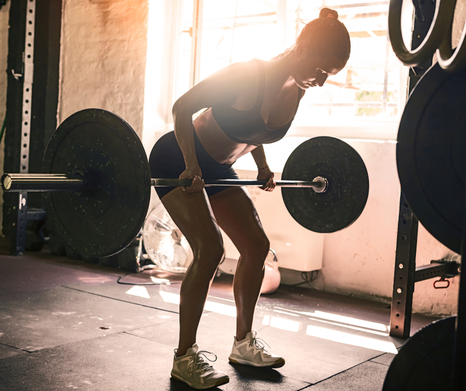
Exercise is essential, but not all exercise affects our bones in the same way. Walking, hiking, and dancing are beneficial weight-bearing activities, while resistance training provides the mechanical loading that helps stimulate bone adaptation. I particularly encourage women to lift weights and progressively challenge their muscles. Strengthening the legs, hips, back, and core also improves the physical capabilities that help prevent falls.
Higher-impact activities can provide additional bone-building stimulus for women who can perform them safely. Some exercise programs use jumping, hopping, or controlled drop landings, such as stepping off a low platform. For women without osteoporosis, vertebral fractures, balance problems, or certain joint conditions, I often recommend jumping from a one-foot box twenty times twice daily. Studies of impact loading support benefits for bone density in selected populations, but this isn’t universally accepted as being enough. I find it easy to remember and could be kind of fun.
I want women to find an exercise program that challenges their bones without creating unnecessary injury risk. For some, that includes jumping. For others, progressive strength training, brisk walking, stair climbing, or carefully supervised impact exercise is a better choice.
Balance and flexibility are equally important because preventing fractures involves preventing falls. Yoga, Pilates, and tai chi can improve aspects of balance, mobility, coordination, and body awareness. Tai chi and structured balance programs have evidence for reducing falls in older adults. Women with established osteoporosis should avoid certain loaded spinal-flexion and twisting movements, particularly if they have vertebral fractures.
The goal is to build a body that is strong enough to support us and coordinated enough to respond when we lose our footing.
Calcium, Vitamin D, Protein, and the Supplements
Nutrition provides the raw materials our bones need to maintain their strength. Women over 50 generally need approximately 1,200 mg of calcium daily from food and supplements combined. I prefer that most calcium come from food because it provides other nutrients alongside it. Dairy products such as yogurt, milk, and cheese are familiar sources, but women who avoid dairy can get calcium from calcium-set tofu, fortified plant milks, canned salmon or sardines with edible bones, kale, bok choy, and other calcium-rich foods.
If dietary calcium is insufficient, supplementation can help fill the gap. Calcium citrate is well absorbed with or without food and is often a good choice for women taking acid-suppressing medications. Calcium carbonate is less expensive and contains more elemental calcium per tablet, but it is best taken with meals. Both can be effective. Because calcium absorption decreases with larger doses, I generally recommend taking no more than 500 to 600 mg of elemental calcium at a time.
Vitamin D helps the intestines absorb calcium and supports normal bone mineralization. Severe vitamin D deficiency can contribute to weak bones and muscle problems. Vitamin D3 is generally effective for raising vitamin D levels, but more is not necessarily better.
I recommend vitamin D3 supplementation of 2,000 IU daily with women who have limited sun exposure or other risk factors for deficiency. Living in Oregon, where winter sunlight is limited, makes this conversation particularly relevant. However, 2,000 IU is not a universal requirement, and higher doses should be based on individual circumstances rather than latitude alone.
Routine vitamin D blood testing is not recommended for every healthy adult because evidence has not shown that population-wide screening improves outcomes. In my practice, I am more inclined to check vitamin D because Oregon is rather rainy. I often find inadequate levels in people not taking supplementation. I recommend Vitamin D-OH levels be between 50-70, while normal is 30. Excessive vitamin D is also problematic since it can cause hypercalcemia, kidney stones, and kidney problems.
Protein is another essential component of bone health. Bone is not simply a calcium structure. It contains a collagen-rich framework, and maintaining adequate muscle mass helps support the skeleton. I generally encourage women in midlife and beyond to consume approximately 1.0 to 1.2 grams of protein per kilogram of body weight daily, with adjustments for activity, medical conditions, and individual needs. Women doing regular resistance training may benefit from higher intake.
Vitamin K2 and Strontium
Vitamin K plays a role in activating proteins involved in bone mineralization, including osteocalcin. Vitamin K2, particularly the MK-7 form, has attracted interest because it may influence calcium metabolism and bone health. Some studies have reported favorable changes in bone-related markers.
Vitamin K2 directs calcium into bones and away from arteries but clinical trials have not established that taking K2 prevents cardiovascular calcification or cardiovascular events. While it is not a proven treatment for osteoporosis or arterial plaque, but I recommend it for prevention.
One supplement that I changed my mind on is Strontium. Strontium makes DXA results appear better than they really are because it affects the way X-rays are absorbed. An apparent improvement in bone density may partly reflect the presence of strontium rather than stronger bone. For these reasons, I no longer routinely recommend strontium supplements.
Hormone Therapy
Estrogen is one of the most effective treatments for preventing the accelerated bone loss associated with menopause. Menopausal hormone therapy helps preserve bone mineral density and reduces fracture risk while treatment continues.
For women experiencing bothersome menopausal symptoms who are appropriate candidates for hormone therapy, bone protection can be an important additional benefit. Timing, age, cardiovascular health, medical history, and individual risk factors all influence whether treatment is appropriate.
I also get questions about testosterone. Testosterone plays a role in skeletal health, and severe testosterone deficiency in men is a recognized risk factor for osteoporosis. In women, however, we do not have convincing clinical evidence that testosterone therapy prevents fractures or meaningfully improves bone density. I do not prescribe testosterone specifically for osteoporosis prevention.
For women who do not want to use systemic estrogen, or who cannot use it, other treatments are available. Raloxifene, also known by the brand name Evista, is a selective estrogen receptor modulator that acts like estrogen in some tissues while blocking estrogen effects in others. It can improve bone density and reduce vertebral fracture risk in postmenopausal women. It also reduces the risk of certain estrogen-receptor-positive breast cancers.
However, raloxifene has not demonstrated a reduction in hip fractures, may worsen hot flashes, and increases the risk of venous blood clots. It is not appropriate for every woman, particularly those with a history of thromboembolism or elevated stroke risk.
For women with established osteoporosis, especially those at high fracture risk, medications such as bisphosphonates or denosumab may be appropriate. Women at very high fracture risk, including some with multiple vertebral fractures, may benefit from bone-building medications such as teriparatide, abaloparatide, or romosozumab, followed by treatment that preserves the newly gained bone.
These medications have different benefits, risks, monitoring requirements, and treatment sequences. I want women to understand that we have effective options, and that the choice should reflect their actual fracture risk rather than their bone density score alone.
Strong Bones Are About More Than Preventing Fractures
When I think about healthy aging, I don't think only about laboratory values, scan results, or preventing disease. I think about what we want our bodies to allow us to do.
I want women to be able to hike, dance, travel, lift their grandchildren, enjoy sex, explore new places, and move through the world with confidence. I want us to remain physically capable and independent for as long as possible.
This requires muscle strength, balance, flexibility, coordination, and the confidence to remain physically active. A woman with good bone density but poor balance and significant muscle weakness can still be at risk for a serious fall. Conversely, someone with osteopenia who builds strength, improves balance, and receives appropriate treatment may meaningfully reduce her risk of injury.
I encourage women to think about bone health as part of their larger physical future. We cannot control every aspect of aging, but we can influence how we prepare our bodies for the decades ahead.
This week, I encourage you to find out where you stand. Consider whether your reproductive history, family history, medications, or medical conditions place you at greater risk for osteoporosis. Talk with your clinician about whether a DXA scan is appropriate. Look at whether you are eating enough calcium and protein, challenging your muscles, and making time for activities that improve your balance.
You don't need to do everything perfectly, and you don't need to wait until you are 65 to begin caring for your bones.
The goal is to build and preserve the strength that allows us to keep living the lives we want.
